 Did you know that muscles, ligaments, cartilage and bone are both sensitive and responsive to mechanical stimulation? What do we mean by that?
This means that in response to stretching, compression or other mechanical stimuli, cells can change the way they behave and tissues can re-organize themselves.
Why is this important?
When horses move, their joints, ligaments, tendons and muscles experience a variety of loading conditions. At times, these conditions can be beneficial for health. For example, when chondrocytes sense mechanical stresses in the range of intensities they perceive as “good”, they happily produce the necessary amounts of molecules to keep the cartilage matrix healthy and resilient. Not only that, but even inflammation in the joint can be potentially reduced when chondrocytes respond to the right amount of mechanical stimulation 4. In other cases, however, the mechanical stresses chondrocytes experience can be too harsh. This in turn can stimulate chondrocytes to produce molecules that break cartilage down, which can ultimately lead to osteoarthritis.
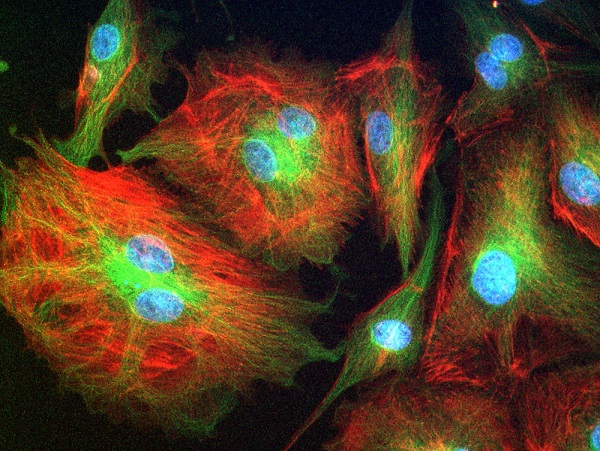
 BPAE cells. Photo by Joseph Elsbernd/flickr/CC BY-NC-SA 2.0. Cells lining the blood vessels, called endothelial cells, can stretch, change shape and divide in response to pressure (blue: cell nuclei; red/green: structures maintaining cell shape).For instance, cells of the cartilage, called chondrocytes, can change in shape due to pressure on the joints during movement, and in response to this mechanical signal can determine whether they need to produce more or less of the cartilage matrix components 1. In response to an exercise program the bones of even very old animals can change their structure and become stronger 2. Similarly, ligaments experiencing a certain degree of tension instruct muscles to contract either more or less 3. These are the examples of how cells and tissues can both sense and respond to mechanical stimuli.
How can we determine whether our horses’ tissues experience excessive amounts of mechanical stresses? Can we learn to recognize movement patterns that create beneficial versus excessive amounts of mechanical stimulation in individual moving horses? Finding the answers to these questions would allow us to prevent injuries, help with injury healing processes and delay the onset and progression of osteoarthritis in our equine partners.
1. te Moller, N. C. R. & van Weeren, P. R. How exercise influences equine joint homeostasis. Vet. J. 222, 60–67 (2017).
2. Leppänen, O. V. et al. Pathogenesis of Age-Related Osteoporosis: Impaired Mechano-Responsiveness of Bone Is Not the Culprit. PLoS One 3, e2540 (2008).
3. Solomonow, M., Hatipkarasulu, S., Zhou, B. H., Baratta, R. V. & Aghazadeh, F. Biomechanics and Electromyography of a Common Idiopathic Low Back Disorder. Spine (Phila. Pa. 1976). 28, 1235–1248 (2003).
4. Ohtsuki, T. et al. Mechanical strain attenuates cytokine-induced ADAMTS9 expression via transient receptor potential vanilloid type 1. Exp. Cell Res. 383, 111556 (2019).
